The different regions of the macula
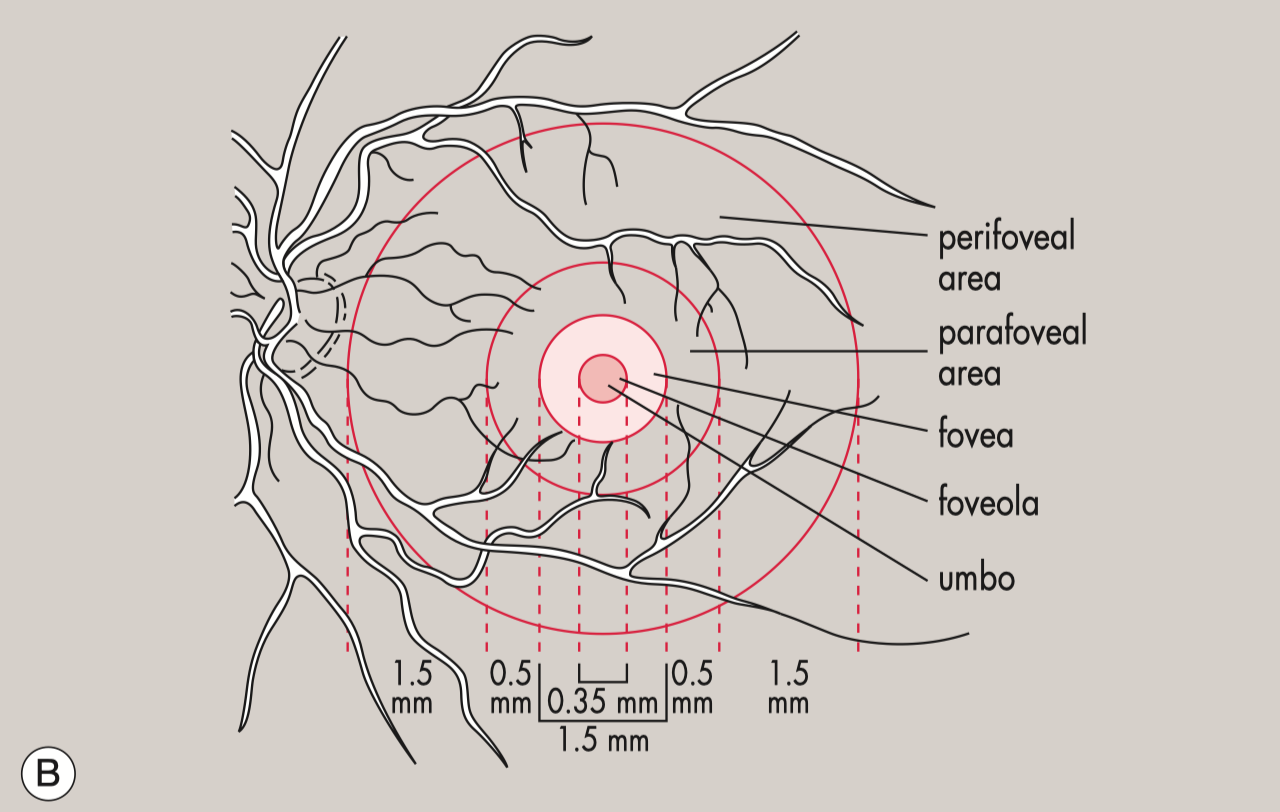
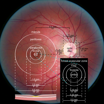
The macular region of eye is made up of the umbo, foveola, fovea, parafoveal and perifoveal.
The macular region of eye is made up of the umbo, foveola, fovea, parafoveal and perifoveal.
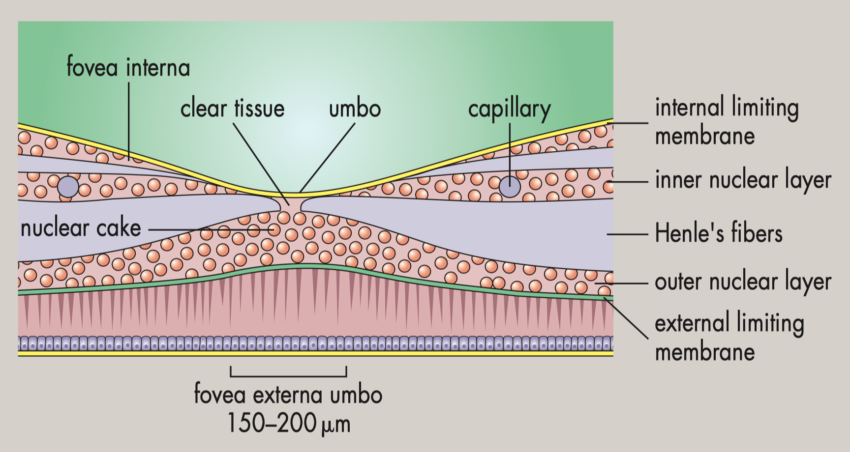
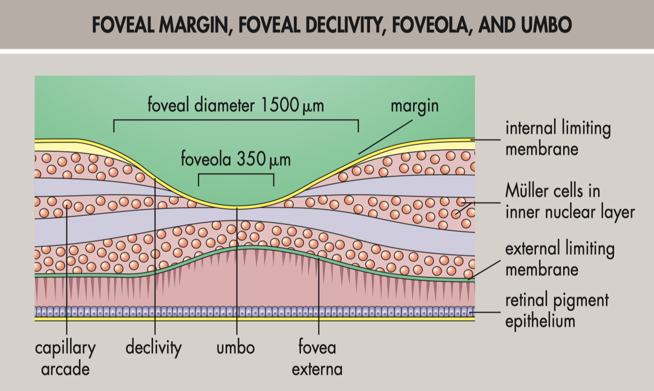
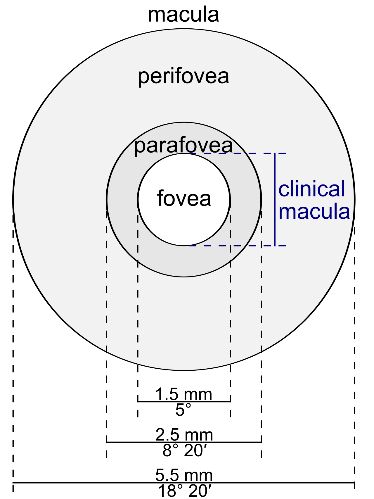
- Umbo is placed at center of fovea's foveola and the foveola is located at center of its fovea.
- The fovea contains the highest concentration of cone cells
- Located in the middle of the fovea, the foveola is roughly 0.35 millimeters in diameter and consists of solely cone cells and a cone-shaped region of Müller cells.
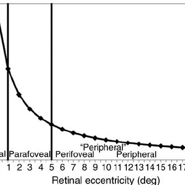
- It has been shown that there are more cone receptors in the foveola than everywhere else in the retina, allowing it to have the best visual clarity.
- Because the cones do not fully form until many months after birth, a newborn's central focus is imperfect.
References
- Anatomy of the Retina: https://www.eophtha.com/posts/anatomy-of-retina.
- Facts and Figures concerning the human retina: https://www.ncbi.nlm.nih.gov/books/NBK11556/
- Wikipedia - Foveola https://en.wikipedia.org/wiki/Foveola
- https://doi.org/10.1001%2Farchopht.117.6.821
- Zeitz, Oliver. "Myron Yanoff and Jay S. Duker: Ophthalmology." (2019)
- Snell, Richard S., and Michael A. Lemp. Clinical anatomy of the eye. John Wiley & Sons, 2013.
- The contributions of central versus peripheral vision to scene gist recognition
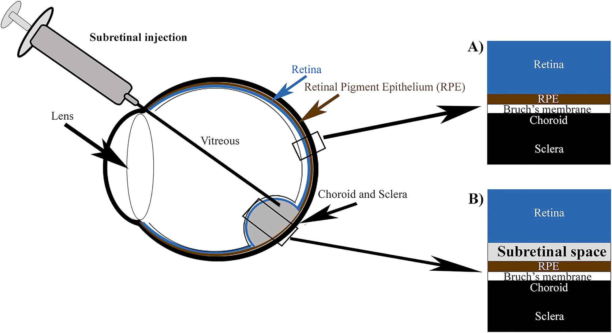
- Subretinal surgery: functional and histological consequences of entry into the subretinal space

Tobiloba Adejumo Newsletter
Join the newsletter to receive the latest updates in your inbox.
